Hot NewsHip Muscles Diagram : Posterior Hip Muscles 800 446 Trailside Fitness
Hip Muscles Diagram : Posterior Hip Muscles 800 446 Trailside Fitness
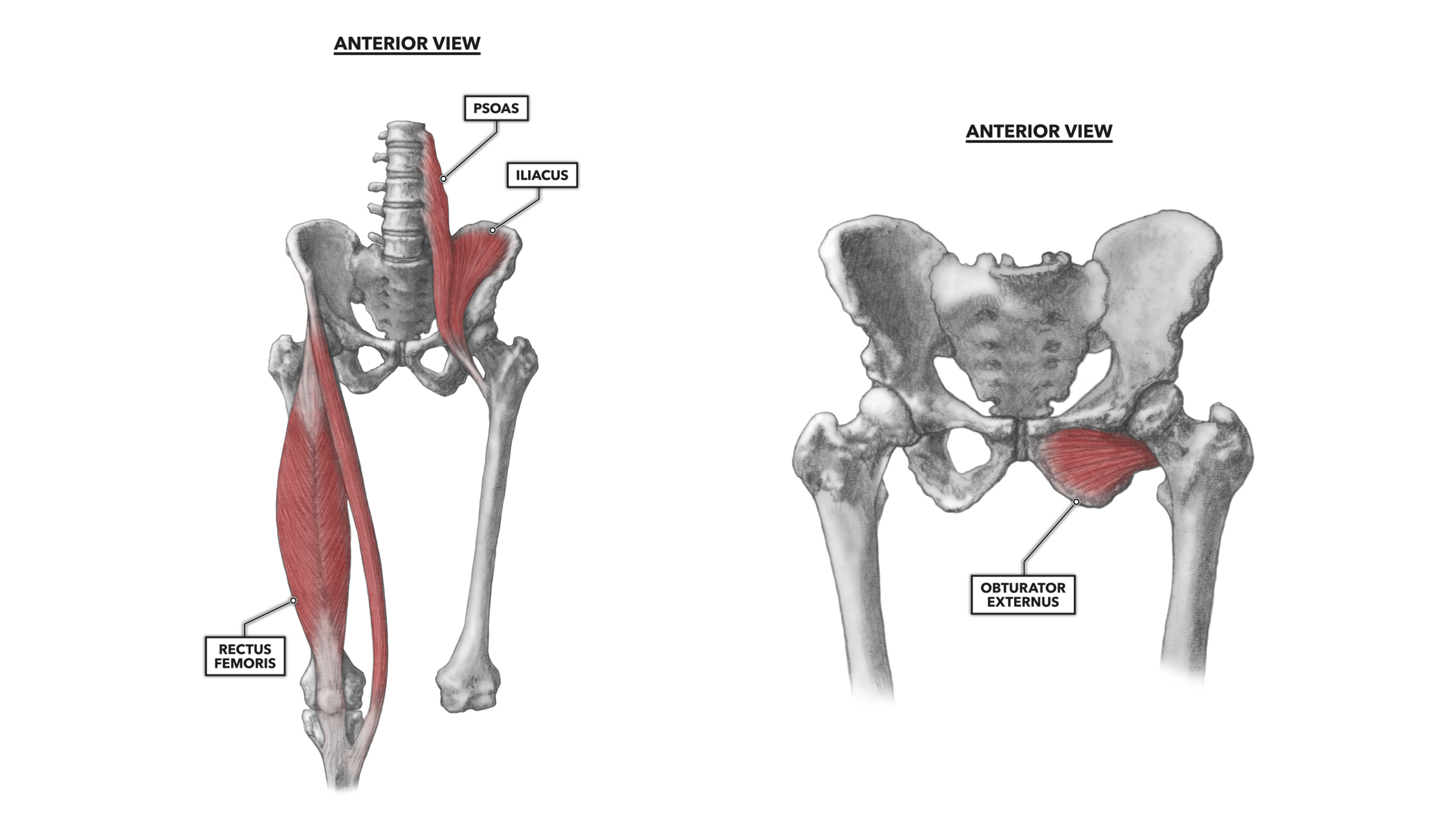
Hip Muscles Diagram : Posterior Hip Muscles 800 446 Trailside Fitness. You can pull your toes up at the exact same time to add another measurement to the stretch. The view on the left has the rectus femoris cut away to show the vastus intermedius which is below it. Muscles play an important role in the. Anatomy of the calf (posterior leg). The hip flexors can be found connecting the top of the femur, which is the largest bone in the body, to the lower back, hips, and groin.
Posted on april 21, 2019april 20, 2019. Rectus femoris muscle, one of the quadriceps muscles on the front of your thigh. There are various hip flexor muscles that all work to. Iliopsoas muscle, a hip flexor muscle that attaches to the upper thigh bone. Extension, flexion, adduction, and abduction.
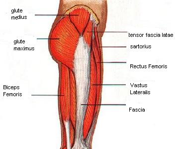
Crossfit Hip Musculature Part 1 Anterior Muscles from www.crossfit.com More commonly, our hips flex to a 90° angle when we sit. You can strain or tear your hip flexor muscles through sudden movements or falls. There are various hip flexor muscles that all work to. As you can see from the diagram to the right, there are many muscles and tendons that make up the hip and buttocks region. Hip muscles diagram an overview of the muscles of the gluteal region, including the superficial and deep gluteal muscles (e.g. Start studying leg/ hip muscles. The diagram shows the posterior (rear) view of the buttock. Diagram of muscles and anatomy charts.
331 615 просмотров • 10 февр.
The view on the left has the rectus femoris cut away to show the vastus intermedius which is below it. Large ligaments, tendons, and muscles around the hip joint hold the bones (ball and socket) in place and keep it from dislocating. Hip pain explained including structures anatomy of the hip and pelvis author juni 02, 2021. Extension, flexion, adduction, and abduction. More commonly, our hips flex to a 90° angle when we sit. When you flex your hip, you move the leg forward. Ligaments, tendons, and muscles play an important role in the function of the hip. Ligaments are soft tissue structures that connect bones to bones.a joint capsule is a watertight sac that surrounds a joint.in the hip, the joint capsule is formed by a group of three strong ligaments that connect the femoral head to the acetabulum. The muscles work together to enable movement and keep the hip in alignment. The hip flexors can be found connecting the top of the femur, which is the largest bone in the body, to the lower back, hips, and groin. Diagram of muscles and anatomy charts. Required to throw a baseball, swing a bat or golf club. The hip flexors are the group of muscles that allow you to lift your knees toward your chest and hip flexor strengthening exercises note:
The diagram shows the posterior (rear) view of the buttock. The hip flexors are the group of muscles that allow you to lift your knees toward your chest and hip flexor strengthening exercises note: Most hip pain stems from limited motion of the hip causing abnormal pressures to different muscles, tendons, or ligaments around the area. The hips are the foundation of our lower bodies. Required to throw a baseball, swing a bat or golf club.
Ligaments Tendons And Muscles Of The Hip Joint Naples Best Hip Surgeon from zehrcenter.b-cdn.net Diagram of muscles and anatomy charts. Iliopsoas muscle, a hip flexor muscle that attaches to the upper thigh bone. These muscles move the thigh toward the body's midline. Learn vocabulary, terms, and more with flashcards, games, and other study tools. The four muscle of the quadriceps all extend the lower leg, and the rectus femoris additionally can flex the thigh at the hip. The muscles in the hip are responsible for the movement of the hip and, by proxy, the leg. Muscles of the lower back and hip diagram hip muscles diagram / click on the labels below to find out more about your muscles. 331 615 просмотров • 10 февр.
Hip pain can sometimes be caused by diseases and conditions in other areas of your body, such as your lower back.
Hip pain on the outside of your hip, upper thigh or outer buttock is usually caused by problems with muscles, ligaments, tendons and other soft tissues that surround your hip joint. This diagram with labels depicts and explains the details of hip muscles diagram. Iliacus, psoas major, psoas minor, obturator externus, obturator internus, superior and inferior gemelli, piriformis, and quadratus femoris muscles. You can strain or tear your hip flexor muscles through sudden movements or falls. Hip pain can sometimes be caused by diseases and conditions in other areas of your body, such as your lower back. Ligaments, tendons, and muscles play an important role in the function of the hip. The view on the left has the rectus femoris cut away to show the vastus intermedius which is below it. The hip flexors can be found connecting the top of the femur, which is the largest bone in the body, to the lower back, hips, and groin. Neck muscle anatomy mri 12 photos of the neck muscle anatomy mri neck muscle anatomy images, neck muscle anatomy pictures, neck muscle anatomy posterior, neck muscle anatomy ultrasound, neck muscles anatomy radiology, human muscles, neck muscle anatomy images, neck muscle anatomy pictures, neck muscle. The muscles in the hip are responsible for the movement of the hip and, by proxy, the leg. The hip muscles encompass many muscles of the hip and thigh whose main function is to act on the thigh at the hip joint and stabilize the pelvis.without them, walking would be impossible. Hip muscles diagram an overview of the muscles of the gluteal region, including the superficial and deep gluteal muscles (e.g. Hip flexion is maximal with a high, forward kick that brings the leg above the level of the waist.
The hip muscles work together to carry out 4 different types of movement: The muscles work together to enable movement and keep the hip in alignment. Start studying leg/ hip muscles. Abducts and rotates thigh laterally, flexes knee at hip, originates at the anterior superior iliac spine and inserts on the medial surface of proximal tibia. Hip pain explained including structures anatomy of the hip and pelvis author juni 02, 2021.
Hip Muscles Pictures And Exercises from www.pilates-back-joint-exercise.com Origin, insertion, action & nerve supply » how to relief. The four muscle of the quadriceps all extend the lower leg, and the rectus femoris additionally can flex the thigh at the hip. The muscles in the hip are responsible for the movement of the hip and, by proxy, the leg. Ligaments are soft tissue structures that connect bones to bones.a joint capsule is a watertight sac that surrounds a joint.in the hip, the joint capsule is formed by a group of three strong ligaments that connect the femoral head to the acetabulum. The hip muscles work together to carry out 4 different types of movement: Large ligaments, tendons, and muscles around the hip joint hold the bones (ball and socket) in place and keep it from dislocating. Neck muscle anatomy mri 12 photos of the neck muscle anatomy mri neck muscle anatomy images, neck muscle anatomy pictures, neck muscle anatomy posterior, neck muscle anatomy ultrasound, neck muscles anatomy radiology, human muscles, neck muscle anatomy images, neck muscle anatomy pictures, neck muscle. Learn vocabulary, terms and more with flashcards, games and other study tools.
Large ligaments, tendons, and muscles around the hip joint hold the bones (ball and socket) in place and keep it from dislocating.
Muscles of the lower back and hip diagram hip muscles diagram / click on the labels below to find out more about your muscles. Snapping hip syndrome is the result of the iliopsoas tendon subluxing over the greater trochanter or ilopectineal eminence. Hip pain on the outside of your hip, upper thigh or outer buttock is usually caused by problems with muscles, ligaments, tendons and other soft tissues that surround your hip joint. Smartdraw includes 1000s of professional healthcare and anatomy chart templates that you can modify and make your own. Diagram of hip muscles and ligaments. They can be divided into three main groups: The hips are the foundation of our lower bodies. The hip muscle diagram below shows a number of the muscles we will be discussing in the next sections. Start studying leg/ hip muscles. Muscles in the human body (pectoralis major, abdominals, obliques). Large ligaments, tendons, and muscles around the hip joint hold the bones (ball and socket) in place and keep it from dislocating. Hip muscles diagram an overview of the muscles of the gluteal region, including the superficial and deep gluteal muscles (e.g. The diagram shows the posterior (rear) view of the buttock.

0 comments